 Fraunhofer Institute for Ceramic Technologies and Systems IKTS
New bone formation after acute inflammation – assessment in vitro
Current research
The group Biological Materials Analysis develops standardized, preclinical test models.
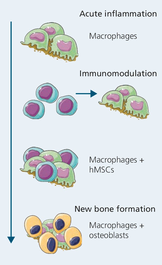
Macrophages and hMSCs interact from acute inflammation until new bone is formed.
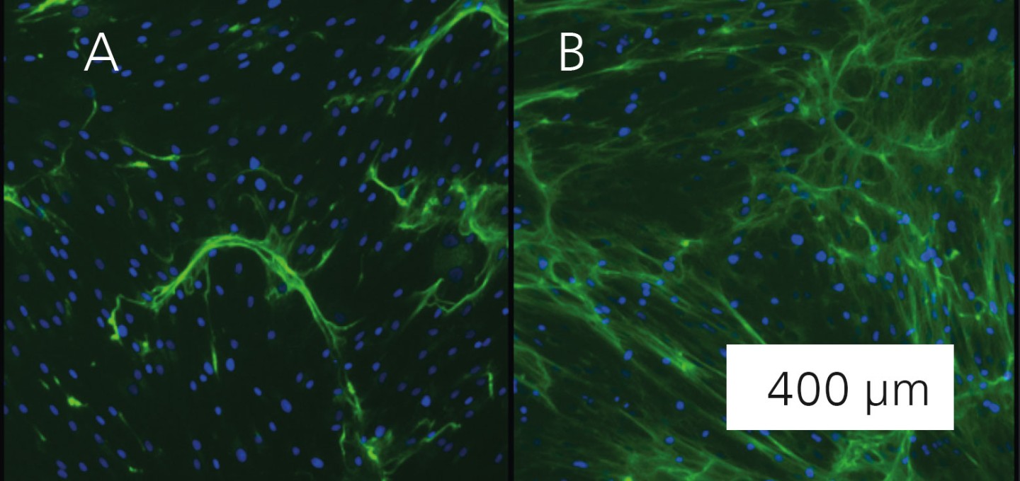
Staining of the generated extracellular matrix (green) via hMSCs without (A) and with inflammatory influence (B). Measuring bar 100 μm.
Whether in bone fractures or the surgical placement of an implant as bone replacement: before the new bone forms, the surrounding tissue is damaged. The immune system responds with acute inflammation at the wound site around the bone.
In the preclinical evaluation of the efficacy of novel implant materials for bone replacement, tests are currently limited to new bone formation. However, it is now known that the preceding acute inflammation has a decisive impact on new bone formation.
Novel osteoimmunology model for preclinical in-vitro testing
The group Biological Materials Analysis at Fraunhofer IKTS has established an in-vitro model that depicts new bone formation under inflammatory conditions. The basis for the combined model consists of two human cell types: macrophages as the protagonists of acute inflammation, and mesenchymal stem cells (hMSCs), which undergo the fundamental process of new bone formation – osteogenic differentiation.
In the model, macrophages are first polarized to a proinflammatory subtype, which creates an inflammatory environment. The process is distinguished by the secretion of proinflam- matory cytokines which, among other symp-toms, leads to redness, swelling and fever in the patient. hMSCs are added to the inflammatory milieu and are subsequently osteogenically differentiated (middle figure). Subsequently, the performance of the osteogenic differentiation is validated and examined based on the production of an extracellular matrix as well as the deposition of calcium (bottom figure). During the investigation of test materials (active substances or [bio]materials), the polarization of macrophages already provides impor-tant insights into the immunomodulatory properties of the test item. Thus, the increase or decrease of inflammation is analyzed as a first step (middle figure, acute inflammation). In addition, the resolution of the inflammation via the immunomodulatory active hMSCs is also characterized (middle figure, immunomodulation). In a second step, the formation of new bone substance is assessed based on the performance of the osteogenic differentiation of the hMSCs under inflammatory conditions (middle figure, new bone formation). The process can be adapted depending on the questions asked about the test item (materials/ active substances).
Analysis portfolio
- Secretion (ELISA, Multiplex)
- Gene expression
- Enzyme activity
- Biochemical assays (e.g., protein-, calcium determination)
- Immunofluorescence staining
Services offered
- Translation of test models onto test items (materials/active substances)
- Assessment of immunomodulatory prop-erties of test materials/-substances
- Assessment of the new bone formation under inflammatory conditions
- Translation of test models for industry, as well as norms and standards