Fraunhofer Institute for Ceramic Technologies and Systems IKTS
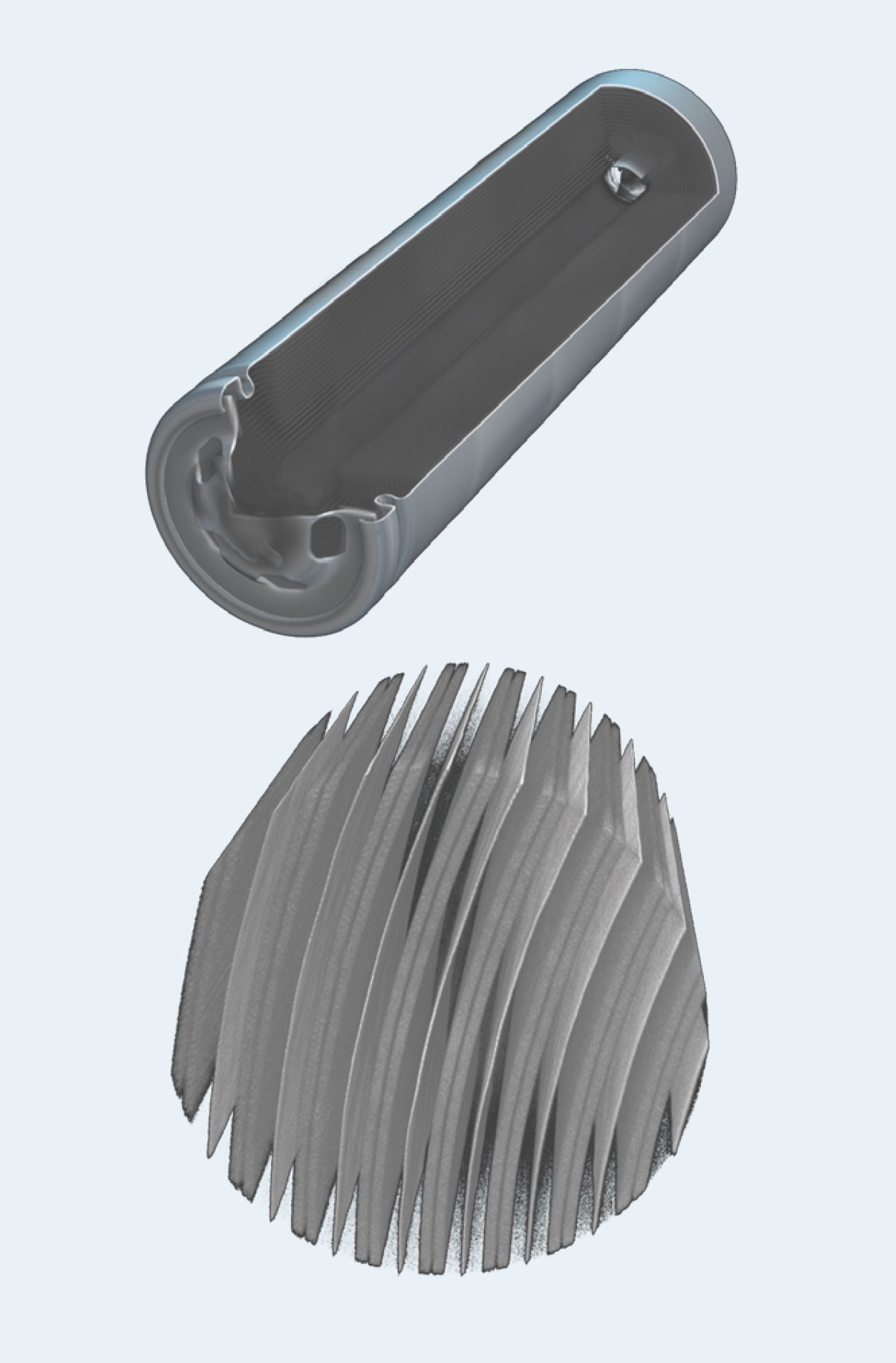
Powerful and safe battery concepts will be essential for a sustainable energy and mobility revolution. The performance of current established technology of the Li-ion battery (LIB) and new concepts, such as all solid-state batteries (ASSB), therefore needs to be continuously improved. Important for this are parameters that can be obtained from multi-modal, crossscale microscopy and spectroscopy, e.g. size distributions of particles in the cathode foil, porosities in the total volume, or interfacial properties of electrode and separator or electrolyte and active material. At its Forchheim site, Fraunhofer IKTS offers precisely this interaction from microscopy to spectroscopy, the correlation of data from different analytical methods from the macro to the nano scale. A fully comprehensive preparative workflow is available for the characterization of battery materials, which avoids oxidation and thermal artifacts while allowing cells and components to be opened and prepared under inert gas and, if necessary, cryogenically cooled during preparation and analysis.
Non-destructive detection of defects
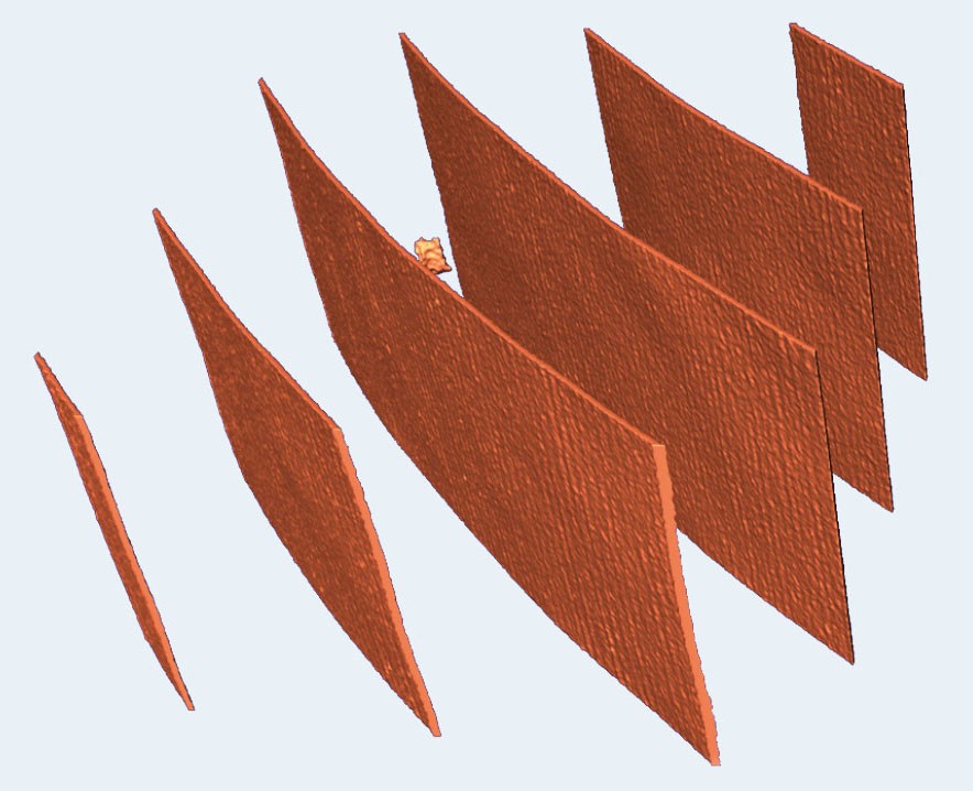
The top figure shows reconstructed X-ray tomography images of a Li-ion battery as an overview and in detailed view. X-ray tomography microscopy (CT with voxel sizes of 500 nm upwards) is able to generate non-destructive three-dimensional images of the internal microstructure of the battery. This method allows to detect structural changes that would otherwise only be visible with invasive methods. A copper outgrowth on the copper current conductor (middle figure) is clearly visible after dedicated image segmentation. Such copper outgrowths occur while the battery is being used and lead to irreparable damage. Such defects can be delayed or avoided by optimizing the material and cell concepts, for which multi-modal analysis can be used.
Cryo-vacuum preparation and dedicated multi-modal, cross-scale, correlated characterization of material, components and integrated devices
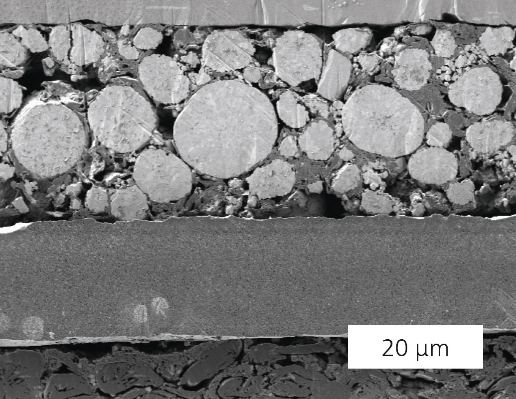
At Fraunhofer IKTS in Forchheim, battery cells, components and starting materials of various types can be opened in an inert atmosphere and transferred to preparation tools or microscopes and spectrometers using an airtight sample shuttle. In order to achieve the highest surface quality, the samples are processed with a cryogenically cooled ion beam polish to prevent the alteration of materials due to heating (e.g. for polymer electrolytes). High-resolution images in the scanning electron microscope (e.g. bottom image), which can also be integrated tomographically from a large number of images to form a 3D volume, provide access to parameters such as particle size, distribution, pore volume, interfacial property, layer thickness and cell structures with a high degree of statistical certainty.
Digital workflow
The entirety of all data from the analytics can be quantitatively evaluated and correlated on an interactive data platform. The data can be used for training machine learning algorithms, e.g. to enable and speed up the automated segmentation of new image data. The resulting database can also be used to generate synthetic data or to set up realistic simulations.