
Nano-Global Positioning System (nanoGPS) for correlative imaging and analytical data acquisition
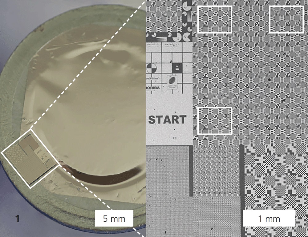
Although exposure to micro- and nanoplastic particles (MNPs) found in food and air is ubiquitous, the potential effects on human health, especially on internal organs, are largely unknown. Accurate risk assessment requires knowledge of the concentration and agglomeration of MNPs in the environment and in physiological media, as well as detection of MNPs using analytical techniques. For the first task, Fraunhofer IKTS has many years of experience and measurement methods; for the second, the newly developed relocalization technology nano-GPSR (Horiba Scientific) is suitable. It is based on a hardware/ software combination and facilitates the use of a correlative microscopy and spectroscopy workflow for the investigation of physical and chemical properties on one and the same object in the nano range. The nanoGPSR tag serves as a position transducer that is firmly attached to the sample under investigation and is moved back and forth with it between different analysis devices (Figure 1). It contains lithographic patterns that are imaged at different resolutions. Software identifies these patterns for a given magnification, allowing the determination of sample coordinates in the different instruments.
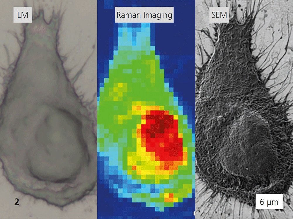
The use of nanoGPSR technology to combine analytical and imaging modalities leads to a comprehensive understanding of the processes in cells (here: podocytes, which act as filter barriers in the kidney) and tissues in relation to plastic exposure. Human kidney cells (Figure 2) were selected to demonstrate the accumulation of MNPs over the lifetime and deterioration of cell health. Micro-Raman spectroscopy is used to characterize the different types of plastics. Cell damage from MNP exposure is inferred from imaging using light and electron microscopy. By overlapping these data, it is possible to avoid overestimation of particle size and underestimation of particle number for clusters and individual MNPs and to obtain Raman measurement signals of MNPs. After incubation of podocytes with four different MNP types, cell viability assays, which determine the proportion of living cells, showed that the decrease in cell viability starts at different concentrations for different polymer types.
Services offered
- nanoGPSR-enabled correlative microscopies/spectroscopies
- Development of application-specific context analytics
- Characterization of MNPs in physiological media
- Weathering of MNPs under marine conditions