 Fraunhofer Institute for Ceramic Technologies and Systems IKTS
High-resolution microscopic and spectroscopic methods for water analysis
Current research
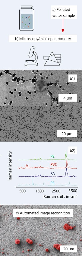
Workflow for water analysis from sampling (a) to filtering (b1) and microscopic (b1) as well as spectroscopic (b2) analysis including automated image recognition using machine learning methods (c).
Water pollution with plastics leads to health hazards worldwide. Macroscopic plastic parts weather in the environment to form micro- and nanoparticles (MNPs), which enter our food chain via plants and animals. The continuous monitoring of the extent of these hazards requires standardized reproducible, artefact-free and ideally automated high-resolution microscopic and spectroscopic methods (Figure a, b). This includes appropriate sample preparation. At Fraunhofer IKTS, these methods are established multimodally and across scales. IKTS has successfully validated its methodology in an international round robin study with 22 laboratories led by the Southern California Water Research Project (SCCWRP) [1]. An important factor for this study was the automated evaluation of the plastic MNPs, e.g. in electron microscopic images based on the training of a U-Net for automated particle detection [2,3] (Figure c). Whether these are plastic MNPs or other types of particles (inorganic, organic) must be determined through additional molecular fingerprinting, e.g. by Raman spectroscopy or infrared spectroscopy (FTIR). Different types of polymers (e.g. PE, PS, PA, PVC, PET and others) can be differentiated in this way as well. Using nanoGPS technology [4], high-resolution microscopy (Figure b1) and spectrometry (Figure b2) can be performed on identical particles with a lateral resolution of ~10 μm (FTIR) or ~1 μm (Raman). The analytical workflow established at Fraunhofer IKTS includes a regulation for water filtration in laminar flow boxes with sieves with mesh sizes from macro to nano scales. This is followed by light microscopy for rapid imaging of entire filters and automated counting and classification of particles up to ~1 μm in size, as well as data correlation with sub-1 μm resolution. Spectral matching with standardized reference libraries (commercial/open source) enables reliable mapping of measured particles.
Recommendations for analyses
Using optical spectroscopy (infrared spectroscopy FTIR) and Raman spectroscopy (Figure b1), particles down to ~1 μm can be recorded automatically with measurement times of < 3 min per particle. Workflows for the analysis of plastic MNPs are also established in complex matrices such as tissues at Fraunhofer IKTS.
Services offered
- Standardized water monitoring and high-resolution microscopic and spectroscopic characterization of plastic MNP residues
- Development of customer-specific preparative and analytical workflows for plastic MNP diagnostics in water samples and more complex matrices, e.g. tissues
- Automated segmentation of plastic MNPs using machine learning
Literature
[1] H. De Frond, G. Sarau, ..., S. Christiansen, Chemosphere, 137300 (2022).[2] Ronneberger, O., Fischer, P., Brox, T. (2015). U-Net: Convolutional Networks for BiomedicalImage Segmentation. In: Medical Image Computing and Computer-Assisted Intervention –MICCAI 2015. https://doi.org/10.1007/978-3-319-24574-4_28. [3] L. Mill, …, S. Christiansen, Small methods 5, 2100223 (2021). [4] G. Sarau, … S. Christiansen, Appl. Spectrosc. 000370282091625 (2020).