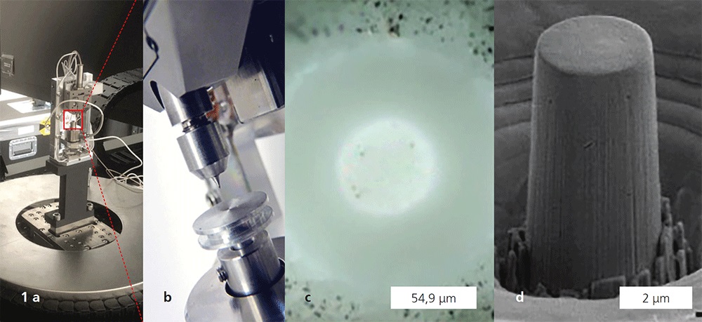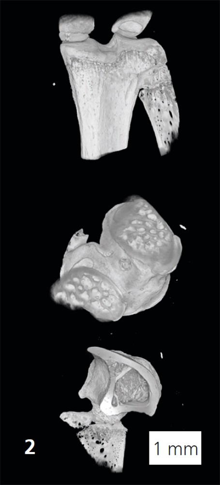

The “4D+ nanoSCOPE” project aims to revolutionize our knowledge of osteoporosis. The six-year joint project funded by the European Union in the module “Excellent Science – ERC-Synergy” brings together three different skill sets: Prof. Georg Schett, Head of Department of Medicine 3 at Universitätsklinikum Erlangen, provides the medical expertise and the sample material. Prof. Andreas Maier, Chair of Pattern Recognition at Friedrich-Alexander-Universität Erlangen-Nürnberg, contributes machine-learning methods to enable an automated and statistically validated interpretation of measurement data. Prof. Silke Christiansen from the Fraunhofer IKTS site Forchheim provides a comprehensive sample analysis consisting of scale-bridging microscopy and spectroscopy to create an unprecedented analytical context. The time-dependent monitoring of bone remodeling dynamics in relation to load and drug therapy represents the fourth dimension, which can only be achieved by in-vivo investigation of bone remodeling (in a mouse model). This requires XRM tomography with the highest resolution and fast image acquisition, which involves new hardware and software developments at the XRM. These are carried out in close cooperation with the company Carl Zeiss. Bone, as a complex and dynamically changing material, has various structural elements such as the compact outer bone wall and a less dense framework architecture in the bone interior. Both structures are interspersed with pore and canal networks that are responsible for bone remodeling and restoration, but at the same time influence the mechanical properties. Biomechanical test procedures on the corresponding length scales (nano to mesoscale) offer the possibility to precisely analyze the mechanical properties, such as tensile and compressive strength, fatigue behavior and fracture toughness, in the different stages of development of osteoporosis and to link them to the bone microstructure. Together with important further analyses, such as Micro-Raman spectrometry, laser scanning microscopy and light sheet fluorescence microscopy, the results are embedded in a correlative-analytical context. This allows the results from the individual procedures to be put together to form a large overall picture, like pieces of a puzzle. Machine-learning methods allow computer-aided detection of recurring patterns in image data. These can be correlated with other diagnostic findings and enable an automated image evaluation with high throughput.
Services offered
- Context microscopy and spectroscopy workflows
- Development of application-specific context analytics
