Fraunhofer Institute for Ceramic Technologies and Systems IKTS
Improved stent properties by laser-induced periodic surface structuring
Current research
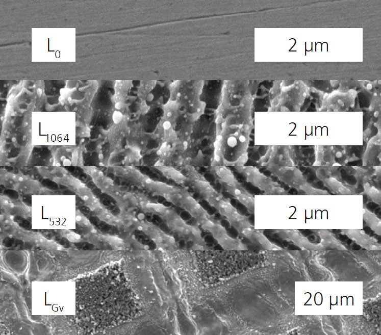
Surface morphology of untreated (Lo) and laser-treated stainless steel samples by picosecond laser system.
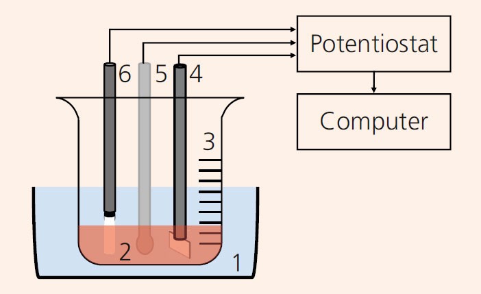
Experimental setup for electrochemical
measurements on stents. 1. Circulating water bath 2. Hank‘s Balanced Salt
Solution (HBSS) 3. Glass beaker 4. Counter electrode: platinum sheet (25*25 mm2), 5. Reference
electrode: saturated
calomel electrode (SCE) 6.
Working electrode: TiON stent.
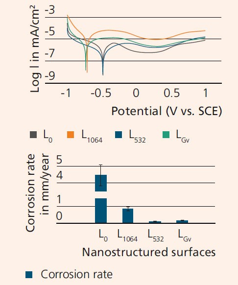
Corrosion behavior of LIPSS
surfaces: Tafel curves (top)
and bar chart for corrosion
rates (bottom).
For several decades, stents have been successfully applied to treat coronary artery disease. Despite this fact, the biocompatibility of stents still needs to be further improved, in particular with regard to antirestenosis, endothelialization and corrosion.
In collaboration with the University of Modena & Reggio Emilia (Italy), NanoPrime (Poland) and the University of Latvia, IKTS researchers investigated the influence of laser-induced periodic surface structuring (LIPSS) on stent properties. This is a promising surface modification technique to influence the migration and proliferation (growth) of cells as well as the corrosion rate of metallic stents.
Three test scenarios
For the tests, AISI 316L stainless steel plates (2 mm in thickness and 10 mm in diameter) were prepared through laser cutting and polishing and were subsequently treated by an EKSPLA Atlantic 5 picosecond laser (beamlines of 1064 nm and 532 nm wavelength). The focus was on three different laser treatments: two, L1064 and L532, aimed to obtain a uniform distribution of LIPSS, while the third, LGv, was supposed to generate a grid of grooves 40 μm in spacing.
Biocompatibility was assessed using human umbilical cord mesenchymal steam cells. The morphology, wettability and structure of the surfaces were evaluated through scanning electron microscopy with energy dispersive X-ray analysis (SEM-EDX), contact angle measurements and laser profilometry.
All LIPSS samples showed increased roughness compared with the reference state and changes of wettability from the hydrophilic to the hydrophobic state. Electrochemical corrosion tests demonstrated the increased corrosion resistance of the laser-treated surfaces associated with their hydrophobic behavior. Among all LIPSS samples tested, the L532 specimen demonstrated the highest corrosion resistance and the highest cell attachment and proliferation with significant influence on cell shape and directional growth along the nanogrooves.
Promising results
The results of this study show that stent surface properties may be enhanced through the application of micro- or nanopatterned coatings or surface modifications. In addition, the treated surface has a positive effect on cellmaterial interaction by suppressing in-stent restenosis and promoting reendothelialization. Since LIPSS structures are capable of mimicking natural nano-properties of vascular tissue, there is great potential for biological transformation.
Sponsored by